Glosarry
Below is a list of common eye-care terms.- Accommodation – The ability of the eye to change its focus from distant to near objects. This is achieved by the lens changing its shape.
- Age-Related Macular Degeneration – Macular Degeneration is a leading cause of vision loss among people over age 50. It results from changes to the macula, a portion of the retina, which is located on the inside back layer of the eye. The macula is responsible for clear, sharp vision and is many times more sensitive than the rest of the retina. Without a healthy macula, seeing detail or vivid color is not possible.
- Amblyopia – A condition of diminished visual acuity in the absence of any detectable cause. It is sometimes referred to as “lazy eye.”
- Astigmatism – A condition that occurs when the front surface of the eye, the cornea, is slightly irregular in shape. This irregular shape prevents light from focusing properly on the back of the eye (the retina). As a result, vision may be blurred at all distances.
- Blind Spot – 1) A small area of the retina where the optic nerve enters the eye; occurs normally in all eyes. 2) Any gap in the visual field corresponding to an area of the retina where no visual cells are present.
- Cataract – A cataract is a clouding of part or the entire lens inside the eye, resulting in blurred or distorted vision.
Color Blindness – The inability to distinguish some colors and shades. It affects about 8% of men and 1% of women in the U.S. - Conjunctiva – The thin, moist tissue that lines the inner surfaces of the eyelids and the outer surface of the sclera (the outer layer of the eyeball).
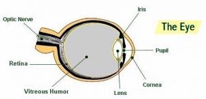
- Cornea – The transparent tissue that covers the front of the eye.
- Farsightedness (Hyperopia) – The inability to see objects up close. It is the result of an eyeball that is too short or an eyeball’s outside surface (the cornea) that is too flat. The exact cause is not known, although farsightedness may be inherited.
- Glaucoma – An eye disease in which the passages that allow fluid in the eye to drain become clogged or blocked, or there is too much fluid produced inside the eye. Increased pressure inside the eye then damages the optic nerve and causes vision loss.
- Intraocular Pressure – Pressure of the fluid inside the eye.
- Iris – The colored ring of tissue suspended behind the cornea and immediately in front of the lens. It regulates the amount of light entering the eye by adjusting the size of the pupil.
- Lens – The clear structure located behind the pupil. Its primary function is to provide fine-tuning for focusing and reading. The lens performs this function by altering its shape.
- Legal Blindness – In the U.S., 1) Visual acuity of 20/200 or worse in the better eye with corrective lenses (20/200 means that a person must be at 20 feet from an eye chart to see what a person with normal vision can see at 200 feet), or 2) Visual field restricted to 20 degrees diameter or less (tunnel vision) in the better eye.
- Macula – The small, sensitive area of the central retina. It provides vision for fine work and reading.
- Nearsightedness (Myopia) – The inability to see clearly at a distance. It is caused by an eyeball that is too long or whose outside surface (the cornea) is too curved. Nearsightedness can be inherited or caused by the stress of concentrating for long periods on work at a close distance.
- Optician – A professional who is trained and licensed to make and fit glasses and contact lenses from your prescription or by analyzing your current glasses.
- Optometrist – An optometrist is a doctor of optometry with special training in examining eyes, prescribing glasses, and recognizing eye diseases.
- Ophthalmologist – An ophthalmologist is a doctor of medicine who specializes in treatment of eye diseases through medication, therapy, or surgery.
 Peripheral Vision – Side vision. The ability to see objects and movement outside of the direct line of vision.
- Presbyopia – The gradual loss of the eye’s ability to change focus for seeing near objects. It happens because, with age, the lens inside the eye gradually loses its flexibility and focusing ability. It occurs in almost all people over age 45.
- Pupil – The adjustable opening at the center of the iris that allows varying amounts of light to enter the eye.
- Retina – The light-sensitive layer of tissue that lines the back of the eyeball. It sends visual impulses through the optic nerve to the brain.
Sclera – The tough, white, outer layer of the eyeball. Along with the cornea, it protects the entire eyeball. - Visual Acuity – The ability to distinguish details and shapes of objects. It is also called central vision.
- Visual Field – The entire area that can be seen when the eye is looking straight ahead, including peripheral side vision.
Providing Buffalo, NY with eye exam services, eyeglass frames & more!
Call or visit one of our four Buffalo Area Locations today!